This case study presents the journey of a patient who came to me after experiencing pain from a faulty filling done by another dentist. The initial treatment was performed without proper examination and only addressed the surface issue for a low fee. As a result, the patient developed severe pain, tenderness, and a periapical infection at the root tip, putting the tooth at serious risk.
Clinical Assessment and Patient Discussion:
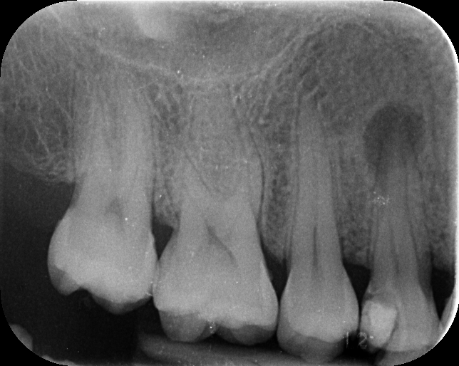
Upon examination and radiographic analysis, I found that the previous dentist had not properly diagnosed the underlying problem. The tooth was nearly lost due to the infection. The patient was very concerned and expressed a strong desire to save the tooth at any cost. I explained that the prognosis was poor, but I would use my best skills to try to save it.
Treatment Steps:
- I recommended root canal therapy. While I usually complete this in a single visit, in this case, I planned a two-visit approach due to the severity of the infection.
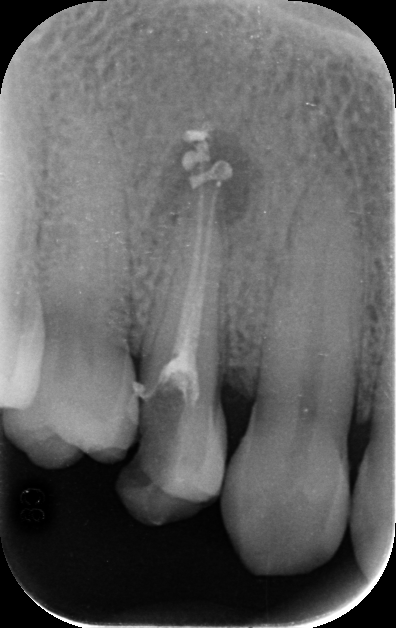
- In the first visit, I placed an intracanal medicament (calcium hydroxide) to help heal the lesion.
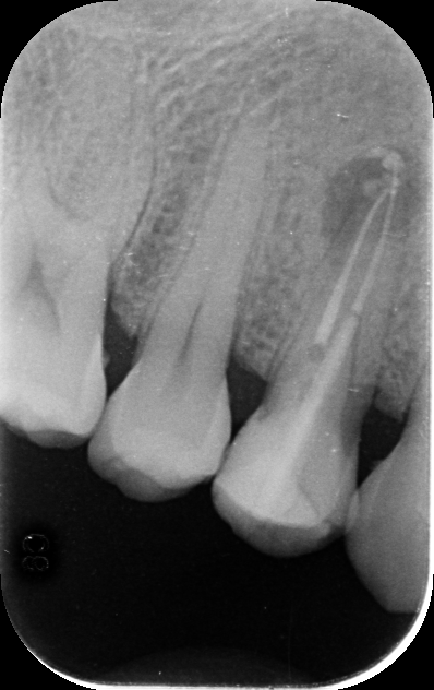
- After 10 days, at the second appointment, I removed the medicament and filled the canals with permanent root canal material.
- I restored the tooth with composite filling and discussed with the patient that placing a crown immediately was not necessary. Instead, I advised waiting one year to assess healing before considering a crown, to avoid unnecessary expenses.
Follow-Up and Results:
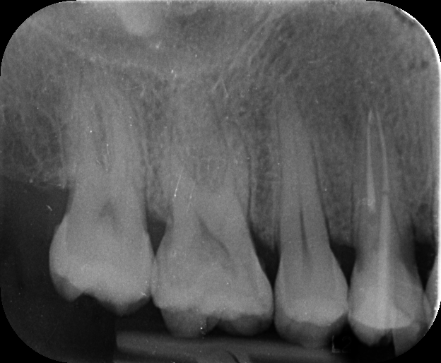
- After one year, the patient returned for a check-up. Clinically, the tooth was healthy, and the fillings were intact.
- A follow-up radiograph showed remarkable healing—about 95% of the lesion had resolved, and the tooth was stable.
- Based on these excellent results, I recommended proceeding with a crown for long-term protection.
Regional Context:
This case reflects a common issue in countries like India, Pakistan, Bangladesh, and neighboring regions, where many people believe simple, low-cost fillings can solve dental problems. Many dentists perform such procedures without proper diagnosis, leading to complications and tooth loss. This case demonstrates the importance of professional care, thorough examination, and patient-centered treatment planning.
Example Note:
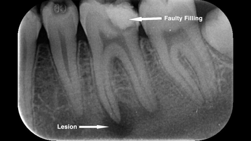
A similar problem can be seen in the case below, where a faulty filling led to the development of a periapical lesion at the root tip. This highlights how inadequate restorations can cause serious complications if the underlying issue is not properly addressed.
About the Author:
This case was managed and documented by Dr. Rouf ul Aziz, Passion Dentistry. All images and content belong to me and my clinic—unauthorized use or reproduction is not permitted.
At Passion Dentistry, we are dedicated to providing high-quality, ethical dental care and sharing knowledge for educational purposes only.
For more information, visit https://www.passiondentistry.com or contact us directly.